StudentAid
Unit 4: Human Urinary System — Long Questions
10th Class Biology · Unit 4: Human Urinary System
Key Points Part of Human urinary system (Kidney, Ureter, Urinary bladder and Urethra)
1. Introduction
The urinary system regulates the internal environment of body. It ensures that waste products and excess substances are removed from body.
2. Part of Human urinary system
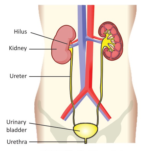
The human urinary system (excretory system) consists of a pair of kidneys, a pair of ureters, a urinary bladder and a urethra.
- Kidney: Kidneys remove extra water, salts and nitrogenous wastes from blood and make urine.
- Ureter: From each kidney, a tube called ureter carries urine to the urinary bladder.
- The urinary bladder: temporarily stores urine.
- The urethra: is the tube that carries urine from the urinary bladder to the outside.
Key Points Shape | Location | Size | Structure of the Kidney (External and Internal Structure)
1. Introduction
Kidneys are the main organs of urinary system. Renal arteries supply them blood with metabolic wastes (e.g., urea, extra water and salts). Kidneys filter these wastes from blood and then remove in the form of urine out of body.
2. Shape
Kidneys are bean-shaped organs having a concave and a convex side. Kidneys are dark-red in colour.
3. Location
They are present in the abdominal cavity, below the diaphragm, on the sides of vertebral column. The concave side of each kidney is towards vertebral column. The left kidney is a little higher than the right.
4. Size
Each kidney is about 10 cm long, 5 cm wide and 4 cm thick.
5. Structure of the Kidney
a. External Structure
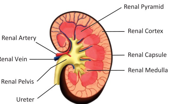
i. Renal Capsule: Kidney is surrounded by a tough tissue called renal capsule.
ii. Hilum: On the concave side of kidney, there is a depression called hilum. It is the point from where renal artery enters in kidney and renal vein and ureter leave the kidney.
b. Internal Structure
i. Renal Cortex & Renal Medulla: Internally, a kidney is divided into two regions. The outer region is called renal cortex while the inner one is called renal medulla.
ii. Renal Pyramids: There are many cone-shaped areas in renal medulla, called renal pyramids.
iii. Renal Pelvis: The base of each pyramid faces the renal cortex while its tip is in a cavity called renal pelvis. It extends to outside of kidneys and forms a ureter.
Key Points Parts of Nephron (Renal Corpuscle and Renal Tubule)
1. Introduction
The functional units of kidneys are called nephrons. There are more than one million nephrons in each kidney.
2. Parts of a Nephron
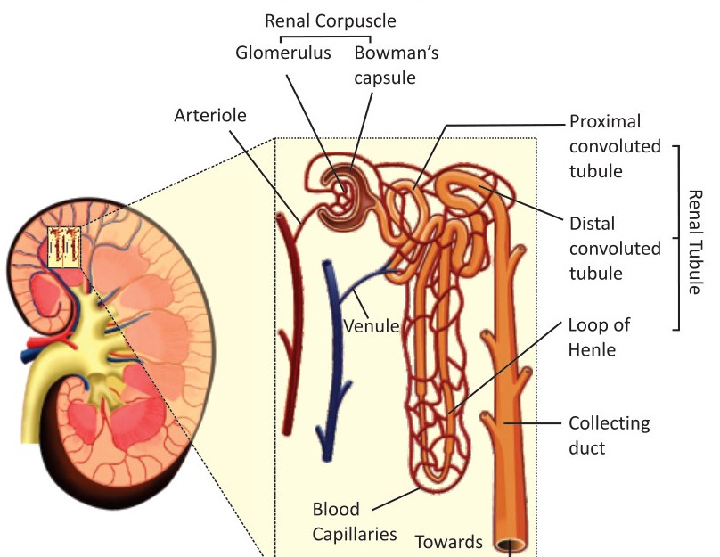
A nephron consists of two parts i.e., renal corpuscle and renal tubule.
a. Renal Corpuscle: It is the first part of nephron and is composed of glomerulus and the Bowman's capsule.
i. Glomerulus: is a network of capillaries.
ii. Bowman's capsule: is the cup-shaped structure around the glomerulus.
b. Renal tubule: It is a long tube attached with the Bowman's capsule. It has following parts:
i. Proximal Convoluted Tubule: The first part is convoluted and is called the proximal convoluted tubule.
ii. Loop of Henle: The middle part is U-shaped and is called the Loop of Henle.
iii. Distal Convoluted Tubule: The last part is again convoluted and is called the distal convoluted tubule.
iv. Collecting Duct: The distal convoluted tubules of many nephrons open in a single collecting duct. Many collecting ducts join together and open into the renal pelvis.
Key Points Pressure filtration | Reabsorption | Tubular Secretion
1. Introduction
Kidneys control the chemical composition of blood. They take extra and waste materials from blood and convert them to urine. It occurs in three steps.
2. Pressure Filtration
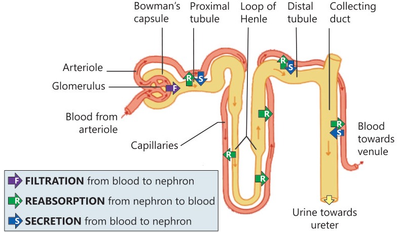
a. Process of Filtration: Blood enters kidney through a renal artery. The artery divides into many arterioles. Each arteriole divides into the capillaries of glomerulus.
When blood reaches glomerulus, its pressure is very high. So, many small molecules (water, salts, glucose and urea etc.) move out of glomerulus. These materials enter Bowman's capsule. This step is called pressure filtration.
b. Glomerular Filtrate: The filtered material in Bowman's capsule is called glomerular filtrate. It is about 20% of the blood plasma that enters the glomerulus.
3. Reabsorption
a. Introduction: The glomerular filtrate moves to the next part of nephron i.e., renal tubule. Many useful materials are reabsorbed from glomerular filtrate into the blood capillaries surrounding the renal tubule.
b. Reabsorption at Proximal Convoluted Tubule: Large amount of water, nearly 100% of glucose and amino acids, and most of the salts are reabsorbed from proximal convoluted tubule.
c. Reabsorption at Loop of Henle: Some water is reabsorbed from descending limb of the loop of Henle. Large amounts of salts are reabsorbed from ascending limb of the loop of Henle.
d. Reabsorption at Distal Convoluted Tubule: Some water is also reabsorbed from distal convoluted tubule and collecting duct.
4. Tubular Secretion / What is tubular secretion? How it helps in the removing wastes from blood?
a. Introduction: When useful materials are reabsorbed into blood, some of the remaining wastes present in blood are secreted from blood capillaries to renal tubule. It is an active transport process and is called tubular secretion.
b. Urine and its Movement: After reabsorption and tubular secretion, the filtrate in the renal tubules is known as urine. Urine from all nephrons moves to the renal pelvis. From renal pelvis, urine moves to the urinary bladder via ureters. Here it is stored. When the urinary bladder is filled, urine is passed out through urethra.
Key Points Formation of Diluted Urine | Formation of Concentrated Urine
1. Introduction
Osmoregulation means the control of the amount of water according to body's needs. Kidneys are the main organs which work for osmoregulation. They control the amount of water in blood. So, the amount of water in other body fluids is also controlled.
2. Formation of Diluted Urine
If there is excess water in blood, more water is filtered from the glomerulus to Bowman's capsule. There is less reabsorption of water from renal tubule to capillaries. In this way, abundant dilute urine is produced and the amount of water in blood is brought to normal.
3. Formation of Concentrated Urine
If the amount of water in blood is less than normal, less water is filtered from the glomerulus to Bowman's capsule. There is more reabsorption of water from renal tubule to capillaries. In this way, concentrated urine is produced and the water is preserved in blood.
Key Points Kidney Stones | Kidney Failure | Treatments of Kidney Disorders
1. Kidney Stones / Write a comprehensive note on kidney stones describing the causes, symptoms and treatment.
a. Introduction: Sometimes, the filtered minerals and salts (e.g., calcium oxalate, calcium and ammonium phosphate, and uric acid etc.) gather in kidneys and make hard, crystalline deposits. Such deposits cannot pass in urine and are called kidney stones. Some stones may leave kidneys and may be trapped in ureter or urinary bladder.
b. Symptoms: Severe pain in kidney or lower abdomen, burning in urethra, frequent urination, foul-smell in urine, blood and pus in urine, nausea, vomiting and bloating.
c. Causes: Presence of more calcium oxalate, calcium and ammonium phosphates in diet (green vegetables, fats, dairy products), extra amounts of vitamins C and D in diet, reduced water intake, excess uric acid in blood, urinary tract infections, and alcohol consumption.
d. Treatment
i. Drinking Plenty of Water: If kidney stones are small in size, the patient is advised to drink plenty of water so that stones can pass through the urine.
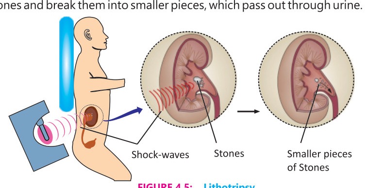
ii. Lithotripsy: is another method for the removal of kidney stones. In lithotripsy, non-electric shock waves are bombarded on kidney from outside. The waves hit the stones and break them into smaller pieces, which pass out through urine.
iii. Surgical Treatment: If stones are large and cannot pass easily, the patient has to undergo surgery for the removal of stones.
2. Kidney (Renal) Failure / Write down the Symptoms and Causes of Kidney Failure
a. Introduction: When one or both kidneys are not able to perform their function (removing waste materials from blood and then passing them out), the condition is called kidney failure.
b. Symptoms: Increases concentration of wastes in blood, rise in volume of body fluids, weight loss, and blood in urine, swelling in legs, feet and face.
c. Causes: Long-term diabetes mellitus, hypertension, dehydration and severe kidney infections, overuse of certain medicines (e.g., nonsteroidal anti-inflammatory drugs- NSAIDs), large kidney stones, physical damage to kidneys, heart or liver failure.
3. Treatment of Kidney Failure / Dialysis / Define Dialysis and Describe the Process of Peritoneal Dialysis and Haemodialysis
a. Introduction: Dialysis is a treatment in which wastes are extracted from blood by using a dialysis fluid. There are two methods of dialysis:
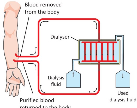
b. Haemodialysis: In haemodialysis, the patient's blood is pumped through a machine called dialyzer.
- The dialyzer contains long tubes. Blood flows inside the tubes while dialysis fluid flows outside.
- Extra water and wastes move from blood and enter the dialysis fluid.
- The purified blood is then returned back to the body.
c. Peritoneal Dialysis
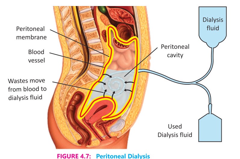
- In this method, dialysis fluid is pumped into the peritoneal cavity (the space around gut in abdomen).
- The walls of peritoneal cavity are lined with peritoneal membrane.
- The wastes pass from the blood present in blood vessels of peritoneal membrane into the dialysis fluid.
- After some time, the dialysis fluid is drained out of the body.
4. Kidney Transplantation
a. Introduction: Kidney transplantation is done for advanced treatment of kidney failure.
b. Conditions for Kidney Transplant
i. In this treatment, damaged kidney of a patient is replaced by a healthy kidney from some donor.
ii. The donor of kidney may be a deceased person or a living one.
iii. Living-donor may or may not be a relative of the patient.
iv. In all cases, the donor's kidney is matched with the patient's immune system.
c. Life of Donated Kidney: When a matching kidney is transplanted, it works normally in patient's body for 10 – 15 years.
More figures from this unit