StudentAid
Unit 3: Human Blood Circulatory System — Long Questions
10th Class Biology · Unit 3: Human Blood Circulatory System
The transport system consists of two key parts i.e.,
(i) Blood circulatory system
(ii) Lymphatic system
The main components of human blood circulatory system are:
• Blood
• Heart
• Blood vessels
Key Points Components of blood | Blood plasma | Blood cells and cell-like body (RBCs, WBCs and Platelets)
1. Introduction: Blood is a type of connective tissue.
2. Functions
• Its main function is to transport materials through the body
• It carries important materials to where they are needed.
• It transports oxygen from the lungs to all body cells and carries carbon dioxide back to the lungs for removal.
• Blood also delivers nutrients from the digestive system to the cells, hormones from glands to target organs, and waste products from the cells to kidneys for excretion.
3. Components of Blood | Describe the different components of blood
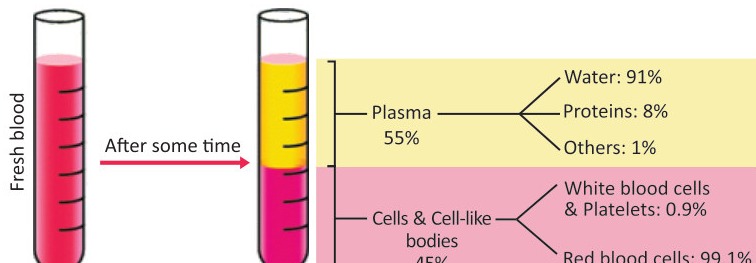
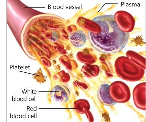
About 55% of blood is composed of a fluid portion called plasma while 45% of blood is made up of cells or cell-like bodies. The adult human has about 5 litres blood in the body.
a. Blood Plasma
i. Introduction: It is the liquid portion of blood. Plasma is composed by 90-92% of water, 7-9% of proteins and 1% of other substances.
ii. Proteins
• Antibodies are important plasma proteins. Antibodies defend the body against pathogens.
• Fibrinogen is also a plasma protein. It is responsible for blood clotting.
• Albumin is a plasma protein which maintains the osmotic pressure of blood.
iii. Salts
• The important plasma salts are made up of sodium, chloride, and bicarbonate ions.
• In addition, there are little amounts of calcium, magnesium, copper, potassium, and zinc.
iv. Nutrients, Wastes and Hormones
• Plasma contains nutrients like glucose, lipids, and amino acids etc. They enter blood from the digestive system.
• The wastes produced by cells are also present in plasma.
• Hormones secreted by endocrine glands are also carried by plasma.
v. Respiratory Gases
• Small amounts of carbon dioxide and oxygen are dissolved in plasma.
• Oxygen is mainly carried by RBCs but about 1.5% of oxygen is also present in plasma in dissolved form.
• Similarly, about 5- 7% of carbon dioxide is carried as dissolved in plasma.
b. Blood Cells and Cell – like Bodies
i. Red Blood Cells (Erythrocytes)
• Structure: Red blood cells (RBCs) are disc-shaped cells with a depression at the centre. They contain a red protein, haemoglobin.
• Functions: It transports oxygen and little amount of carbon dioxide
• Number: There are 4 to 5.5 million RBCs per mm³ of blood.
• Formation: Before and immediately after birth, RBCs are formed in liver and spleen. In adults, they are formed in the red bone marrow of short bones (sternum, ribs and vertebrae). During the formation of an RBCs, its nucleus and organelles are broken down.
• Life Span: The average life span of an RBC is about 120 days.
• Removal of RBCs: When RBCs complete their age, they are removed by spleen, bone marrow, and liver.
ii. White Blood Cells (Leukocytes)
• Introduction: White blood cells (WBCs) are colourless and have irregular shapes.
• Formation: They are formed in the red bone marrow. Some WBCs mature in lymph nodes, tonsils, thymus gland, or spleen.
• Size and Number: WBCs are larger in size and less in number than RBCs. Each mm³ of blood normally contains about 7,000 WBCs.
• Functions: They defend the body against diseases.
• Life Span: Their life span depends upon their needs.
• Types of White Blood Cells
There are several types of WBCs.
(a) Granulocytes
i. Introduction: These WBCs have granules in cytoplasm. They include neutrophils, eosinophils, and basophils. Their names tell the staining properties of their cytoplasm.
ii. Types
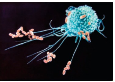
• Neutrophils destroy bacteria and clean up dead cells at infection sites.
• Eosinophils fight with parasites and cause allergic reactions.
• Basophils release histamine to cause inflammation and produce allergic responses.
(b) Agranulocytes
i. Introduction: They have clear cytoplasm. There are two types of agranulocytes i.e., monocytes and lymphocytes.
ii. Types
• Monocytes make macrophages which engulf the germs and dead cells.
• Lymphocytes make antibodies against pathogens.
iii. Cell-like Bodies i.e., Platelets (Thrombocytes)
• Introduction: Platelets are not whole cells. They are small fragments of large cells of bone marrow. Platelets lack a nucleus.
• Life Span: Their life span is 7 to 12 days.
• Number: A mm³ of blood contains 250,000 platelets.
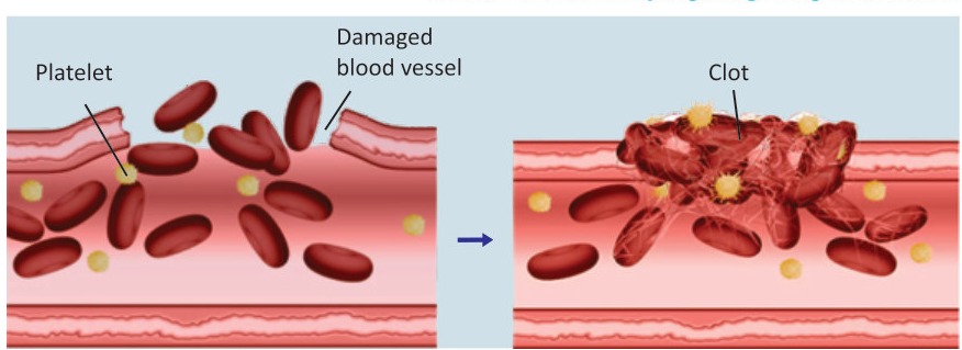
• Functions: When a blood vessel is damaged, platelets gather at the damaged site. Here, they convert plasma protein fibrinogen into fibrin. The fibrin molecules form a net that traps RBCs. The mass of fibrin and RBCs hardens. This hard mass is called a clot. The clot prevents bleeding until the damaged vessel is repaired.
Key Points Location | Structure of Heart (Pericardium, septum, chambers and valves)
1. Introduction
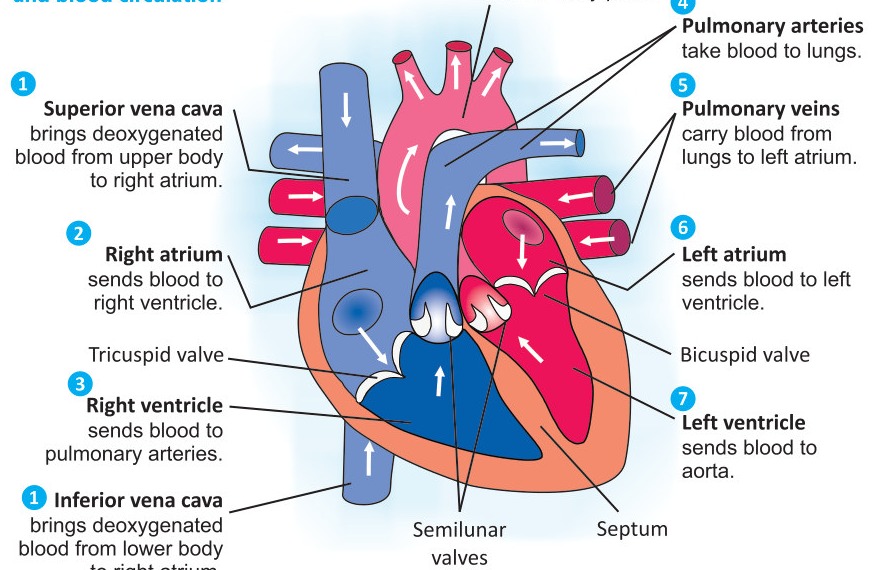
The heart is a muscular organ that pumps blood through a network of blood vessels.
2. Location
The heart lies within the chest cavity, behind the breast bone (Sternum), between the two lungs.
3. Structure of the Heart
a. Pericardium: There is a tough, sac-like membrane around the heart. It is called pericardium. It secretes a fluid around heart. This fluid reduces friction between the pericardium and heart.
b. Septum: Like birds and other mammals, human heart has two sides. The wall between the left and right sides is called septum.
c. Chambers: Each side is divided into two chambers. The upper chambers are called atria (singular: atrium), and lower chambers are called ventricles. The atria have thinner walls as compared to ventricles.
d. Heart Valves
Special flaps called valves are present between the chambers of both sides. The valves open in only one direction.
i. Tricuspid Valve: The valve between right atrium and right ventricle is called tricuspid valve (made of 3 flaps).
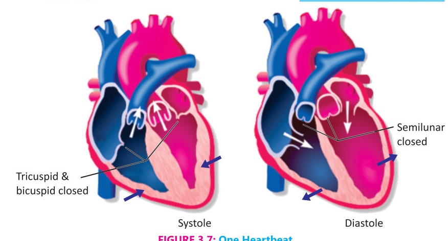
ii. Bicuspid Valve: The valve between left atrium and left ventricle is called bicuspid valve (made of 2 flaps). As the ventricles contract, tricuspid and bicuspid valves close. So, the blood cannot flow back into atria. In this way, blood is pumped from the ventricles into large blood vessels.
iii. Semilunar Valve: A semilunar valve is present between each ventricles and large blood vessels. These valves prevent blood from flowing back into the ventricles.
Key Points Heat as double pump | Circulation of deoxygenated and oxygenated blood | Pulmonary and systemic circulations
1. The Circulation of Blood
a. Working of Human Heart as a Double Pump
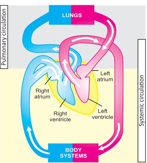
The right side of the heart collects blood from body and sends it to the lungs while the left side collects blood from the lungs and sends it to the body. It means that human heart works as a double pump. It is done in the following way.
b. Circulation of Deoxygenated Blood
Two veins i.e., superior and inferior vena cava bring deoxygenated blood (with high concentration of CO₂ and low concentration of O₂) from parts of the body (other than lungs). These veins open in the right atrium. The right atrium contracts and sends this blood into the right ventricle. The right ventricle contracts and pumps this blood into pulmonary arteries. The pulmonary arteries take this blood to lungs. In lungs, CO₂ diffuses out of the blood, and O₂ diffuses into the blood.
c. Circulation of Oxygenated Blood
From lungs, pulmonary veins carry the oxygenated blood back to heart. These veins open in the left atrium of heart. Then blood is pumped into left ventricle.
When left ventricle contracts it pumps the blood into a large blood vessel called aorta. From aorta, this blood is transported to all parts of the body.
2. Pulmonary and Systemic Circulations
The flow of blood from heart to lungs and then from heart is called pulmonary circulation. Similarly, the flow of blood from heart to the body tissues and then from body tissues to heart is called systemic circulation.
Key Points Heartbeat | Average human heartbeat | Phases of heartbeat
1. Heartbeat
The alternating contraction (systole) and relaxation (diastole) of heart chambers makes one heartbeat.
2. Average Human Heartbeat
The average human heart beats 70 times per minute. This is also called the heart rate.
3. Phases of Heartbeat
a. Systole occurs when both ventricles contract to pump blood into the pulmonary arteries and aorta.
b. Diastole occurs immediately after systole when both atria relax so that blood enters the atria. Contraction of the atria fills the ventricles.
Key Points Arteries | Formation of arterioles and capillaries | Capillaries | Formation of venules and veins | Veins
1. Arteries
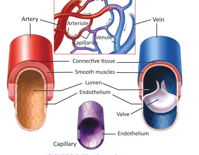
a. Introduction: Arteries carry blood away from the heart. In adults, all arteries carry oxygenated blood, with the exception of pulmonary arteries.
b. Structure
Arteries are strong and elastic. The hollow internal cavity of arteries in which the blood flows is called lumen.
The thick walls of arteries are made up of:
• An inner layer of endothelium.
• A middle layer of smooth muscles and elastic tissues.
• An outer layer of connective tissue.
c. Formation of Arterioles and Capillaries
When arteries enter body organs, they divide into smaller vessels known as arterioles. The arterioles enter tissues and divide into capillaries.
2. Capillaries
a. Introduction: Capillaries are the smallest blood vessels.
b. Structure: The walls of capillaries are composed of only a single layer of cells i.e., endothelium. This layer is so thin that water, nutrients and oxygen can pass through it to enter the tissue fluid. Similarly, carbon dioxide and other wastes present in tissue fluid can pass through it to enter blood.
c. Formation of Venules and Veins
In tissues, capillaries unite to form small veins, called venules. The venules unite to form veins.
3. Veins
a. Introduction: Veins carry blood towards the heart. In adults, all veins carry deoxygenated blood, with the exception of pulmonary veins.
b. Structure
The walls of vein are composed of the same three layers as are present in the artery wall i.e.
(i) An inner layer of endothelium.
(ii) A middle layer of smooth muscle and elastic tissues. In veins, the middle layer is comparatively thin as compared to artery. It has lesser smooth muscles and elastic tissues.
(iii) An outer layer of connective tissue.
(iv) The lumen of the veins is broader than that of arteries.
(v) Most veins have valves that prevent the backflow of blood.
Key Points Arterial system | Arteries of pulmonary circulation | Arteries of Systemic Circulation
Arterial System
1. Arteries of Pulmonary Circulation
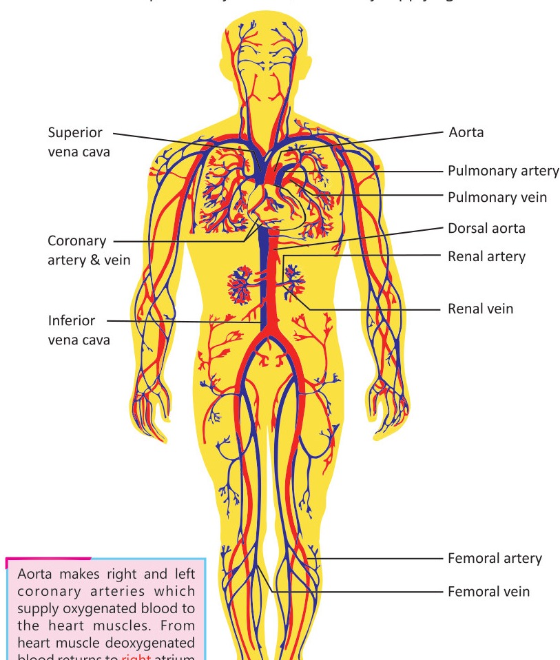
a. Pulmonary Trunk: A large artery called pulmonary trunk carries deoxygenated blood from right ventricle.
b. Pulmonary Arteries: It branches into two smaller pulmonary arteries, with one artery going to each lung.
2. Arteries of Systemic Circulation
a. Aorta: Oxygenated blood is pumped from left ventricle into aorta. It forms arteries which supply blood to head, shoulders and arms.
b. Dorsal Aorta: Aorta passes through the thorax and becomes dorsal aorta. It makes many arteries that supply blood to all parts of the lower region. For example,
(i) Hepatic Artery supplies blood to the liver.
(ii) Renal Arteries supply blood to kidneys.
(iii) Femoral Arteries: Aorta divides and makes two femoral arteries which supply blood to legs.
Key Points Veins of Pulmonary Circulation | Veins of Systemic Circulation
Venous System
1. Veins of Pulmonary Circulation
Two pulmonary veins carry oxygenated blood from lungs to the left atrium of heart.
2. Veins of Systemic Circulation
a. Two major veins i.e., superior vena cava and the inferior vena cava carry deoxygenated blood from body to right atrium.
b. The superior vena cava is made by joining of veins from head, shoulders and arms.
c. The inferior vena cava is made up of many veins from parts of the lower region. For example:
(i) Femoral Veins: Two femoral veins from legs empty into inferior vena cava.
(ii) Renal Veins: Renal veins carry blood from the kidneys.
(iii) Hepatic Vein: From liver, a hepatic vein carries blood to the inferior vena cava.
Key Points Causes | Symptoms | Risk factor | Prevention | Complications
1. Introduction
The diseases that involve the heart or blood vessels are collectively called cardiovascular disorders or cardiovascular diseases (CVDs).
2. Coronary Heart Disease (CHD)
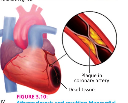
It is the narrowing or blockage of the coronary arteries, which supply blood to the heart muscle. This condition is primarily caused by atherosclerosis i.e., accumulation of fatty deposits (plaques) inside arteries.
Causes High levels of low-density lipoproteins (e.g., cholesterol) in blood, high blood pressure, smoking, diabetes, sedentary lifestyle
a. Symptoms: Asymptomatic in early stages, may lead to chest pain or discomfort as the condition progresses.
b. Risk Factors: Family history, obesity, high blood pressure, high cholesterol levels, and lifestyle factors.
c. Prevention: Healthy diet, regular exercise, smoking cessation, and management of blood pressure and cholesterol levels.
d. Complications: If left untreated, CHD can lead to more severe conditions, including myocardial infarction and heart failure.
3. Myocardial Infarction
a. Introduction: "Myocardium" means heart muscle and "Infarction" means tissue death. The death of heart muscles is called myocardial infarction. It is commonly known as a "heart attack". It occurs when blood flow to a part of the heart muscle is blocked for a prolonged period, leading to death of the heart muscle.
b. Causes: A blockage in one or more coronary arteries that forms over atherosclerotic plaques.
c. Symptoms: Chest pain, tightness, and pressure shortness of breath, sweating and pain radiating to the arm, neck, or jaw.
d. Treatment: Emergency treatment involves medicines to dissolve clots, angioplasty to open blocked arteries, and stenting to keep arteries open.
e. Complications: Heart failure, arrhythmias (irregular heartbeats), and sudden cardiac arrest.
f. Prevention: Maintaining weight with a healthy diet, avoiding smoking, exercising regularly, controlling blood pressure, cholesterol and diabetes.
4. Angina Pectoris
a. Introduction: Angina Pectoris (Commonly called Angina) is chest pain or discomfort caused by reduced blood flow to the heart muscle, typically due to coronary artery disease. Angina is a symptom of CHD and often indicates that the heart is not getting enough blood.
b. Symptoms: Chest pain or discomfort, chest pressure and squeezing or fullness. Pain can radiate to the shoulders, neck, or arms.
c. Treatment: Lifestyle changes, medications, procedures like angioplasty or stent placement.
d. Complications: If untreated, can progress to myocardial infarction and increase the risk of heart failure.
Key Points Increase of risk of atherosclerosis, Blood Clot Formation, | Elevated blood pressure | Arrhythmias | Reduced oxygen delivery | Impact on overall cardiovascular health
Harmful effects of smoking related to heart diseases
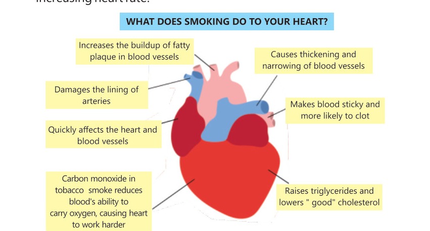
Smoking is the major risk factor for the development of heart diseases. Here's an overview of how smoking adversely impacts heart health.
1. Increased Risk of Atherosclerosis
Smoking accelerates atherosclerosis by damaging the endothelium of arteries.
2. Increased Risk of Blood Clot Formation
Smoking increases the chances for blood to clot by promoting platelets aggregation. The clots that can block coronary arteries and lead to myocardial infarction.
3. Elevated Blood Pressure
Smoking contributes to elevated blood pressure by causing vasoconstriction.
4. Heart Rate and Rhythm Abnormalities
Arrhythmias are irregular heartbeats that can be dangerous. Smoking affects heart rhythm by increasing heart rate.
5. Reduced Oxygen Delivery
Carbon monoxide from cigarette smoke binds to haemoglobin in RBCs more effectively than oxygen, reducing the blood's oxygen-carrying capacity.
6. Impact on Overall Cardiovascular Health
Smoking contributes to the narrowing of arteries in the legs, which can further strain the heart.
More figures from this unit