StudentAid
Unit 5: Coordination — Long Questions
10th Class Biology · Unit 5: Coordination
Key Points Stimulus | Receptor | Coordinator | Effector | Response
Coordinated Action
A coordinated action has five components:
1. Stimulus
a. Definition: Any factor in external or internal environment. which can initiate a response in the body. is called a stimulus.
b. Examples: Touch, light, sound, heat, cold, pressure and infection etc are the stimuli.
2. Receptor
a. Definition: The organ, tissue or cell which receives stimulus is called the receptor.
b. Example: Ears are the receptors for the stimulus of sound. Receptors send the messages of stimulus to coordinators.
3. Coordinator
a. Definition: A coordinator receives information from receptors and sends messages to particular organs for proper action.
b. Examples: In nervous coordination, brain and spinal cord are coordinators. In chemical coordination, endocrine glands are the coordinators.
4. Effector
a. Definition: These are the parts of the coordinated action which receive coordinator's messages and perform specific actions.
b. Examples: In nervous coordination, muscles and glands are the effectors. While in chemical coordination, different tissues of the body act as effectors.
5. Response
a. Definition: A response is the action performed by effectors. on receiving the message from coordinator.
b. Example: Pulling our hand away from something very hot.
Key Points Coordination | Types
1. Coordination: Coordination means integration among different parts of the body and their response to stimuli in order to keep harmony with the environment. Coordination occurs in all multicellular organisms.
2. Systems for Coordination: Animals possess two systems for this function i.e., nervous system and endocrine system.
Key Points Neuron / Nerve Cell | Nerve Impulse | Structure of Neuron
1. Neuron / Nerve Cell: Neurons or nerve cells are the cells that transmit messages in the body in the form of nerve impulses.
2. Nerve Impulse: It is a wave of electrochemical changes that travels across the membranes of neurons.
3. Structure of Neuron: A neuron consists of three major parts i.e., cell body, dendrites and axon.
a. Cell Body: The cell body contains the nucleus, other organelles and most of the cytoplasm.
b. Dendrites: Small branches which project from the cell body are called dendrites. They transmit nerve impulses towards the cell body
c. Axon: Axon is a long branch from cell body. Its terminal end is further branched. Axon transmits nerve impulses from cell body to other neurons, muscles, or glands.
Following structures are associated with neurons;
4. Neuroglia: Neurons are supported by specialized cells, called neuroglia. Schwann cells: Schwann cells are a type of neuroglia.
Key Points Synapse | Neurotransmitters | Function of Neurotransmitters
1. Synapse: A synapse is a small gap between two neurons or between a neuron and an effector cell. When nerve impulse reaches the end of a neuron, it cannot jump directly to the next neuron or effector. It is transmitted with the help of neurotransmitters.
2. Neurotransmitters
Neurotransmitters are special chemicals that help to carry messages across a synapse.
3. Function of Neurotransmitters
When a nerve impulse reaches the end of a neuron, it releases neurotransmitters. They travel across the synapse and attach to the next neuron or effector cell. It helps the nerve impulse continue its journey.
4. Examples
Examples of neurotransmitters include dopamine and serotonin.
Key Points Sensory Neurons | Motor Neurons | Inter-Neurons
Types of Neurons
1. Sensory Neurons transmit nerve impulses from receptors to the CNS.
2. Inter-neurons are present in brain and spinal cord. They receive impulses from sensory neurons, interpret them and transmit them to motor neurons.
3. Motor neurons transmit nerve impulses from inter-neurons to effectors (muscle or glands).
Key Points Nerve | Sensory Nerves | Motor Nerves | Mixed Nerves
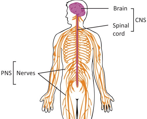
1. Nerve: A nerve is collection of axons that are enveloped by a covering of connective tissue. Nerves arise from brain and spinal cord and make the Peripheral Nervous System (PNS).
2. Types: There are three types of nerves, on the basis of axons present in them.
i. Sensory Nerves: Sensory nerves contain the axons of sensory neurons only.
ii. Motor Nerves: Motor nerves contain the axons of motor neurons only.
iii. Mixed nerves: Mixed nerves contain the axons of sensory and motor neurons.
Key Points CNS | PNS | Composition of Nervous System
1. Introduction
There are two major components of nervous system i.e., central nervous system (CNS) and peripheral nervous system (PNS).
a. CNS: The CNS comprises of brain and spinal cord.
b. PNS: The PNS consists of nerves that arise from brain and spinal cord and spread in different parts of body.
2. Composition of Nervous System
All these components are made of neurons and supporting (Neuroglial cells) cells.
Key Points Brain and its parts (Forebrain, Midbrain, Hindbrain)
The central nervous system consists of brain and spinal cord.
A. Brain / Write the Major Functions of the Forebrain, Midbrain and Hindbrain
1. Introduction
All life activities are under the control of brain. Brain is present inside cranium (part of skull).
2. Meninges: Inside cranium, brain is covered by three layers of membranes, collectively called meninges. Meninges protect the brain from harmful substances.
3. Ventricles: Brain contains fluid-filled cavities called ventricles. The ventricles of brain are continuous with the central canal of spinal cord.
4. Cerebrospinal fluid: Fluid within ventricles and central canal of spinal cord is called cerebrospinal fluid (CSF).
5. Parts of Brain: The brain can be divided into three major parts:
a. Forebrain b. Midbrain c. Hindbrain
a. Forebrain
It is the largest area of brain. Following are the major parts of this region.
i. Cerebrum
• Introduction: It is the largest part of forebrain.
• Cerebral Hemisphere: A deep groove (longitudinal fissure) divides the cerebrum into right and left cerebral hemispheres.
• Cerebral Cortex
The upper layer of both hemispheres is highly folded and is called cerebral cortex. There are two regions in cerebral cortex.
o Grey matter: Containing cell bodies and non-myelinated axons.
o White matter: The material beneath cortex is white matter (containing myelinated axons).
• Lobe of Cerebral Hemisphere | Write a note on lobes of cerebral hemispheres and also discuss their functions.
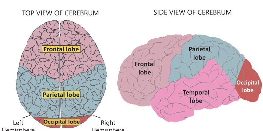
Each hemisphere is further divided into four lobes.
o Frontal Lobe: The frontal lobe contains regions for problem solving, judgment, speaking, and movements.
o Parietal Lobe: The parietal lobe has regions for language and touch.
o Temporal Lobe: The temporal lobe contains regions which deal with memory, learning, feelings, and hearing.
o Occipital Lobe: The occipital lobe has regions for vision and color perception.
ii. Thalamus
• Introduction: It is a structure wrapped by the cerebrum.
• Functions: It conveys information coming from receptors to the cerebrum. Thalamus is also involved in pain perception and consciousness.
iii. Hypothalamus
• Location: It lies above midbrain and just below thalamus.
• Size: In humans, it is about the size of an almond.
• Functions: It regulates body temperature, hunger, thirst, sleep, and emotional states. It also controls the secretions of the major endocrine gland i.e., pituitary gland.
b. Midbrain
i. Location: The midbrain is located between forebrain and hindbrain.
ii. Functions
• It is a bridge between forebrain and hindbrain.
• It conveys information between.
• It also enables the body to make adjustments for movements.
• It is also responsible for sleep/wake, alertness and temperature regulation.
c. Hindbrain
Hindbrain controls all automatic body functions. It consists of three parts.
(i) Medulla oblongata
• Location: It is on the top of the spinal cord.
• Functions
o It transmits impulses between spinal cord and the higher parts of brain.
o It controls autonomic functions such as heartbeat, heart rate, breathing and blood pressure.
o It also controls reflexes such as vomiting, coughing, sneezing.
(ii) Cerebellum
• Location: It is behind medulla oblongata. It is the second largest part. Its surface is highly folded.
• Functions: It coordinates balance and muscle movements.
(iii) Pons
• Location: It is present on top of the medulla.
• Functions: It makes connections between the spinal cord and cerebellum, and also between cerebrum and cerebellum. It also regulates alertness, sleep, and wakefulness.
Key Points Location | Structure | Functions | Spinal Nerves
1. Introduction: It is a tubular bundle of nerves that starts from brain and extends to lower back.
2. Location: It is located in vertebral column.
3. Protection: It is also covered by meninges.
4. Functions
a. Spinal cord transmits impulses between brain and body parts.
b. It also acts as a coordinating centre for some simple reflexes.
5. Structure
a. White Matter: The outer region of spinal cord is made of white matter
b. Grey Matter: The central region is made of grey matter. It is butterfly shaped and surrounds the central canal.
6. Spinal Nerves: Thirty-one pairs of spinal nerves arise from spinal cord. All the spinal nerves are "mixed" nerves because each contains axons of both sensory and motor neurons.
a. Dorsal Root: The dorsal root contains sensory axons and a ganglion where cell bodies are located.
b. Ventral Root: The ventral root contains axons of motor neurons.
Key Points Nerves Pathways | Sensory and Motor Pathways, Somatic Nervous System | Autonomic Nervous System
1. Introduction: It is composed of nerves and their ganglia. Humans have 12 pairs of cranial nerves and 31 pairs of spinal nerves. Some cranial nerves are sensory, some are motor and some are mixed. On the other hand, all spinal nerves are mixed.
2. Nerves Pathways: The cranial and spinal nerves make two pathways;
(a) Sensory Pathway: Conducting impulses from receptors to CNS.
(b) Motor Pathway: Conducting impulse from CNS to effectors. Motor pathway makes two systems.
i. Somatic Nervous System: It controls conscious and voluntary actions. It includes all of the motor neurons that are connected to skeletal muscles.
ii. Autonomic Nervous System: It consists of motor neurons that are connected to cardiac muscles, smooth muscle and glands. It is generally without conscious control. Autonomic nervous system consists of sympathetic and parasympathetic systems.
• Sympathetic Nervous System: Sympathetic nervous system prepares body in emergency situations. This is often called the "fight or flight" response. This system dilates pupils, speeds up heartbeat, increases breathing rate and inhibits digestion.
• Parasympathetic Nervous System: When there is no stress, parasympathetic nervous system normalizes the activity of body. It is called "rest and digest" response. It causes pupils to contract, promotes digestion, and normalizes the rate of heartbeat.
Key Points
Reflex Action | Reflex Arc | Examples
1. Reflex Action: Reflex actions are the quick involuntary response in which brain is not involved. Reflex actions are controlled by a reflex arc.
2. Reflex Arc: A reflex arc is the pathway of neurons over which the nerve impulses travel during a reflex action.
3. Example: We pull our hand away from a hot flame without thinking about it. It is a reflex action and happens in the following way;
• Skin receptors detect the stimulus of heat.
• Sensory neuron sends nerve impulse to inter-neuron of spinal cord.
• The inter-neuron transmits the nerve impulse to motor neuron.
• The motor neuron carries the nerve impulse to effector (muscles of arm).
• The effector produces a response by contracting. So, our hand is pulled.
Key Points
Hormones | Major Endocrine Glands (Pituitary Gland, Thyroid Gland, Parathyroid Gland, Adrenal Gland, Pancreas, Gonads)
1. Introduction
The endocrine system consists of special endocrine glands which produce and secrete hormones for coordination in animals.
2. Hormones
Hormones are the chemicals, released from endocrine gland, which regulate different body functions like growth, reproduction, and glucose level in blood etc.
3. Ductless glands
The endocrine glands do not have ducts to pour their hormones. That is why: they are also called ductless glands. They secrete hormones into blood which carries them to different parts of the body.
4. Major Endocrine glands
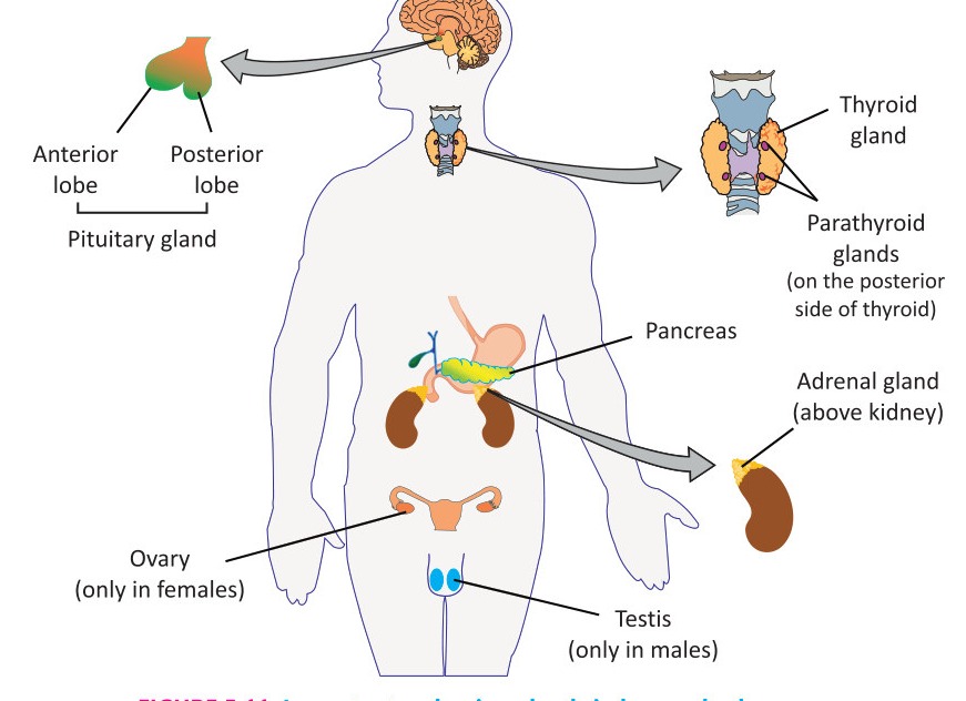
Following are the major endocrine glands and their important hormones.
I. Pituitary Gland / State the Hormones of the Pituitary Gland and Write their Functions
a. Introduction: It is a small gland present in the brain.
b. Location: It is suspended from the hypothalamus by a short stalk.
c. Lobes of Pituitary Gland: There are two lobes of pituitary gland i.e., anterior lobe and posterior lobe.
i. Hormones of Anterior Lobe of Pituitary
• Growth Hormone (GH): It stimulates the growth of muscles, bones, and other tissues.
○ Gigantism: If growth hormone is produced more than normal, it results in extra growth in body. This condition is called gigantism.
○ Dwarfism: If growth hormone is produced less than normal, it results in less growth in body. This condition is known as dwarfism.
• Adreno-Cortico-Tropic Hormone (ACTH) stimulates the adrenal cortex gland to produce its hormones.
• Thyroid-Stimulating Hormone (TSH) stimulates the thyroid gland to produce its hormone (thyroxine).
• Luteinizing Hormone (LH) is needed for the release of egg cells from ovary (ovulation) in females. In males, it stimulates the testes to produce sex hormones.
• Follicle Stimulating Hormone (FSH) Anterior lobe also produces follicle stimulating hormone (FSH) that is involved in the reproductive system.
ii. Hormones of Posterior Lobe of Pituitary
The hypothalamus produces two hormones that are stored in the posterior lobe of pituitary gland.
° Antidiuretic Hormone (ADH), also called vasopressin, increases the reabsorption of water from nephrons in kidneys.
• Oxytocin initiates contractions in uterus during childbirth. It also stimulates the flow of milk during breastfeeding.
II. Thyroid gland
a. Location: It is located near the lower part of the larynx.
b. Hormones: It secretes two hormones.
i. Thyroxine stimulates enzymes of cellular metabolism (glucose oxidation) for energy production. It increases basal metabolic rate and so produces heat.
ii. Calcitonin It stimulates the transfer of calcium ions from blood to bone, where the calcium ions can be used to generate bone tissue. In this way, calcitonin acts to decrease blood calcium levels.
III. Parathyroid Glands
a. Location: The four parathyroid glands are embedded in the back of the thyroid gland.
b. Hormone: They secrete a hormone called parathormone.
c. Functions of Parathormone: It stimulates the transfer of calcium ions from the bones to the blood. Thus, it has the opposite effect of calcitonin.
IV. Adrenal Glands
a. Location: An adrenal gland is located above each kidney.
b. Parts: Each adrenal gland has an inner core, the medulla, and an outer layer, the cortex.
i. Hormones of Adrenal Medulla:
The adrenal medulla produces two hormones: epinephrine (adrenaline) and nor-epinephrine (nor-adrenaline).
• Functions
○ These hormones take part in the emergency response of the sympathetic nervous system.
○ When a person is in emergency, the medulla releases its hormones to prepare the body for "fight-or-flight" response.
○ These hormones increase heart rate, blood pressure, blood glucose level, and blood flow to the heart and lungs.
○ They also stimulate enlargement of the bronchioles and dilation of the pupils.
ii. Hormones of Adrenal Cortex
The adrenal cortex secretes hormones cortisol and aldosterone.
• Functions
○ Cortisol promotes the production of glucose from proteins.
○ Aldosterone raises blood pressure and volume by stimulating salt and water retention by the kidneys.
V. Pancreas
Pancreas has two functions (Acts as exocrine and endocrine Gland):
a. Function as an Exocrine Gland
It is a part of digestive system, where it plays the role of a ducted (exocrine) gland and secretes digestive enzymes.
b. Function as an Endocrine Gland
i. Islets of Langerhans: It also has special cells called the Islets of Langerhans (discovered in 1869 by a German student, Paul Langerhans). These cells function as an endocrine gland.
ii. Hormones: They secrete two hormones i.e., insulin and glucagon. They regulate the level of glucose in the blood.
• Insulin: Insulin lowers the blood glucose level by stimulating body cells to store glucose or use it for energy.
• Glucagon: Glucagon stimulates release of glucose from liver into the bloodstream.
iii. Diabetes Mellitus: In diabetes mellitus, the cells are unable to obtain glucose resulting in high blood glucose concentrations.
• Type-1: In type-1 diabetes mellitus, the pancreas does not produce required insulin. Such patients are treated with daily injections of insulin.
• Type-2: In type-2 diabetes, insulin is produced but the target cells do not respond to it. This type of diabetes can be controlled through exercise and diet.
VI. Gonads
a. Introduction: Gonads (ovaries in females and testes in males) are gamete-producing organs. They also produce sex hormones.
b. Hormones of Gonads
• Estrogen and Progesterone: In females, the ovaries secrete hormones called estrogen and progesterone. These hormones cause the release of egg from the ovary and regulate female secondary sex characteristics (e.g., development of breast).
• Androgens: In males, the testes secrete a group of sex hormones called androgens (e.g., testosterone). It regulates male secondary sex characteristics (e.g., growth of hair on face and coarseness of voice etc.)
Key Points
Table 12.2 Summary of Major Endocrine Glands and their Hormones
Glands | Hormones | Functions
Anterior lobe of pituitary | Growth hormone, Adrenocorticotropic hormone, Thyroid stimulating hormone, Luteinizing hormone | Stimulates the growth of muscles and bones, Stimulates adrenal cortex to secrete hormones, Stimulates the thyroid gland to secrete hormone, Stimulates the ovary to release egg, Stimulates the testes to produce sex hormones
Posterior lobe of pituitary | Antidiuretic oxytocin | Increases the reabsorption of water from nephrons, Stimulates contractions in uterus during childbirth, Stimulates the flow of milk from mother's breasts
Thyroid | Thyroxine, Calcitonin | Stimulates enzymes of cellular metabolism, decreases blood calcium level
Parathyroid | Parathormone | Increases blood calcium level
Adrenal medulla | Epinephrine and nor-epinephrine | Prepare the body for "fight-or-flight" response in emergency
Adrenal cortex | Cortisol, Aldosterone | Promotes production of glucose from proteins, raises blood pressure and volume
Pancreas | Insulin, Glucagon | Lowers blood glucose level, Increases blood glucose level
Ovaries | Estrogen and Progesterone | Cause the release of egg from the ovary and regulate female secondary sex characteristics
Testes | Androgens (Testosterone) | Regulate male secondary sex characteristics
More figures from this unit