StudentAid
Unit 4: Biomolecules — Long Questions
11th Class Biology · Unit 4: Biomolecules
Definition Biochemistry is the study of chemical components and chemical processes, occurring in living organism.
Importance of studying biochemistry All structures of living organisms have biochemical organization and all functions occurring in them are due to biochemical processes taking place in this organization.
• Therefore, a basic knowledge of biochemistry is helpful to understand anatomy and physiology of living organisms. Photosynthesis, respiration, digestion, contraction etc. can be described in biochemical terms.
Chemical composition of protoplasm
• The molecules synthesized by cells and containing carbon are known as organic molecules. They occur naturally only in the bodies of living organisms or in their products and remains.
• Carbohydrates, proteins, lipids and nucleic acids are important organic molecules in living organisms.
• Inorganic molecules: Life on Earth evolved in water, and all life still depends on water At least 80% of the mass of living organisms (protoplasm) is water, and almost all chemical reactions of life take place in aqueous solutions.
• The other chemicals that make up living things are mostly organic macromolecules and certain inorganic molecules.
• Dry mass composition of biomolecules: They make 93% of the dry mass of living organisms. The remaining 7% comprises of small organic molecules (like vitamins) and inorganic molecules (like carbon dioxide, acids, bases, and salts).
Different types of bonds and interactions play vital roles in the structure and function of biological molecules.
Importance of carbon Carbon is the basic element of organic molecules. It is tetravalent and can react with many other elements like H, O, N, P and S. Carbon bond (C-H bond) is the potential source of chemical energy for cellular activities.
• Carbon-oxygen association in glycosidic linkages provides stability to the complex carbohydrate molecules.
• Carbon combines with nitrogen in amino acid linkages to form peptide bonds and forms proteins which are very important due to their diversity in structure and functions.
Bonds
1. Covalent Bonds: Covalent bonds form when two atoms share electrons.
Significance These bonds are often found in organic molecules like proteins and nucleic acids, providing stability to the molecules.
2. Ionic Bonds: Ionic bonds are formed when one atom donates an electron (becomes a positive ion, or cation) and another atom accepts the electron (becomes a negative ion, or anion). The electrostatic attraction between these oppositely charged ions forms the ionic bond.
Significance Ionic bonds are relatively strong in the solid state and are formed mostly in inorganic molecules like sodium chloride.
3. Hydrogen Bond: Hydrogen bonds are weak attractions that occur between a hydrogen atom and an electronegative atom (such as oxygen or nitrogen).
Significance These bonds are important in maintaining the structure of large molecules like proteins and nucleic acids, as well as in various biological processes like DNA replication.
4. Hydrophobic Interactions: Hydrophobic interactions occur between nonpolar molecules and polar molecules (like water). Nonpolar molecules tend to cluster together in aqueous environments to minimize contact with water molecules.
Significance This phenomenon is crucial for the folding of proteins and the formation of lipid bilayers in cell membrane.
5. Hydrophilic Interactions: Hydrophilic interactions occur between polar molecules and water molecules.
Significance These interactions are essential for the dissolution of polar and ionic compounds in water. These interactions help in various biological processes such as nutrient transport and chemical reactions within cells.
Condensation
• All the biomolecules join in their monomers by condensation or dehydration process.
• During condensation, an -OH group is removed from one monomer and a-H atom is removed from another monomer.
• It is also known as dehydration synthesis because the removal of OH and H groups means the removal of a water molecule.
★ Need of energy: Energy is required to break chemical bonds when water is extracted from monomers. So, cells must supply energy to make macromolecules.
Example
• The formation of maltose by two glucose monomers is an example of a condensation reaction.
Hydrolysis
• Hydrolysis is a chemical process in which macromolecule (polymer) is broken down into smaller fragments by the addition of water molecules.
• It is the reverse of dehydration synthesis.
• Cells break bonds between monomers by adding water to them.
• In this process, OH group from a water molecule joins to one monomer and hydrogen joins to the second monomer.
★ This breakdown of macromolecules is essential in various biological processes, such as digestion and cellular respiration, where smaller molecules are needed for energy production.
Example
• Breakdown of maltose into two glucose monomers by the addition of a water molecule is an example of hydrolysis.
Water is an oxide of hydrogen having chemical formula H2O. This seemingly simple molecule has many surprising properties, which give it the status of "the medium of life".
• About two third of our bodies are composed of water and we cannot exist without it.
• In fact, it is the most abundant compound found in all organisms.
• Its concentration varies from 65 to 89 percent in different organisms.
1. Solvent Properties
• The ability of water to dissolve a wide variety of substances is due to its two properties, the polarity of water molecules and the ability of water molecules to form hydrogen bonds.
★ Significance: Hydrogen bonds help in maintaining the three-dimensional structures of proteins and the double helix structure of DNA.
• The water molecule has distinct ends, each with a partial charge. Hydrogen atom is partially positive and oxygen atom is partially negative. Such molecules are called polar molecules.
• Partial negative charge at one end of a water molecule is attracted to partial positive of another water molecule. This weak attraction is called a hydrogen bond. Water forms a network of such bonds. Many of the properties of water are due to hydrogen bonds in water.
• Charged or polar molecules are soluble in water. In the state of solution, ions and water molecules can react with each other easily. So water provides a medium for metabolism.
2. Hydrophobic Exclusion
Non-polar or uncharged molecules are insoluble in water because water molecules do not make hydrogen bonds with them. When they are placed in water, water molecules move them out. The insoluble molecules make hydrophobic associations with one another
For example, lipids molecules are insoluble in water When they are excluded from water, they make strong associations among themselves. Therefore, lipids help to maintain membranes of cells.
3. Heat Capacity
Definition Specific heat capacity is defined as the number of calories (amount of heat) required to raise the temperature of one gram of a substance from 15°C to 16°C (i.e.,1°C).
Specific heat capacity of water: Water has a high specific heat capacity i.e. 4.184 Joules.. It means that water has great ability to absorb and releasing heat with minimum change in its own temperature. Most of the heat energy absorbed by water is used to break hydrogen bonds between its molecules.
Significance Due to this breakage of hydrogen bonds, individual water molecules start moving more freely and temperature of water rises.
Temperature Stabilizer Due to high specific heat capacity, water heats up more slowly. Similarly, when it is given a cooler environment, it holds its temperature longer Water thus works as temperature stabilizer not only for organisms' internal environment but also for their external environment.
4. Heat of Vaporization
Definition It is the amount of heat required to change a liquid to gas.
Specific heat of vaporization of water: Water has high heat of vaporization. So, it absorbs much heat while changing from liquid state to gas. Its heat of vaporization is 574 Kcal/kg which means a considerable amount of heat energy (574 Kcal) is required to change 1kg of liquid water into vapours.
Significance (Cooling Effect)
• Due to heat of vaporization of water, Earth's temperature is kept moderate for the sustenance of life.
• Heat of vaporization of water also provides cooling effects to plants when they transpire and animals when they perspire (sweat).
• Every gram of water that evaporates from plant or animals' body surface removes 574 calories of heat from the body.
5. Cohesion
Definition The force of attraction between same type of molecules is called cohesion. Hydrogen bonds among water molecules enable them to "stick together"
Significance Inside water, molecules have high cohesion.
• The cohesion of water is important for living world. Plants depend on cohesion among water molecules for the transport of water and nutrients from roots to leaves.
• The evaporation of water from a leaf exerts a pulling force on water within xylem vessels of the leaf. Because of this cohesion, the force is relayed through xylem vessels all the way down to roots. As a result, water rises against the force of gravity. Hydrogen bonds also giber water high surface tension. Water behaves as if it were coated with some invisible film. The insect water strider walks on water without breaking surface.
6. Ionization of Water
• When the covalent bonds among the atoms of water molecule break, water is ionized to form hydrogen ions (H⁺) and hydroxyl ions (OH⁻). At normal conditions, this reaction is reversible and equilibrium is maintained.
• At room temperature (25°C), in a litre of water one molecule out of each 550 million is ionized and thus the concentration of each of H⁺ and OH⁻ in pure water remains at 10⁻⁷ moles/litre.
★ Acids combine with OH⁻ ions, leaving H⁺ ions in medium and make medium acidic. Similarly, bases combine with H⁺ ions, leaving OH⁻ ions in medium, and make medium basic.
Significance H⁺ and OH⁻ ions take part in many chemical reactions in the cells e.g., hydrolysis of macromolecules. Relative concentrations of H⁺ and OH⁻ ions determine the acidity and alkalinity of medium i.e., pH of medium. The pH affects the biochemical reactions. Enzymes work best at specific pH.
7. Density of water
• Water exhibits its maximum density at 4°C. Its density decreases when the temperature lowers. It is because of the hydrogen bonds which keep water molecules relatively far apart.
Significance In rivers, streams or lakes, ice is formed on the surface water due to falling of temperature. As ice is less dense than water, it floats on surface. It acts as an insulator and does not allow heat to escape from the water beneath it. In this way aquatic organisms are protected.
Properties of water and benefits to life
Properties | Bonding | Benefits to life
--- | --- | ---
Best Solvent | Polarity | Provides medium for chemical reactions
Maximum heat capacity | Hydrogen bonding | Keeps temperature constant internally and externally for organism
Maximum density at 4°C | Change in hydrogen bonding | Ice floats on water
High heat of vaporization | Hydrogen bonding | Moderates Earth's temperature
Ionization | Covalent bond breaks | Determine the acidity and alkalinity of medium
Cohesion | Polarity, Hydrogen bonding | Water and nutrients are transported from roots to leaves
Carbohydrates
Carbohydrate are naturally occurring organic compounds. The word "carbohydrate" literally means "hydrated carbon". Carbohydrates are synthesized as the primary products of photosynthesis. During photosynthesis, when reduction of CO₂ occurs, the resulting carbohydrate molecule contains carbon, hydrogen and oxygen in the molar ratio of 1:2:1.
Empirical formula Their empirical formula is C(H2O)n where 'n' is the number of carbon atoms.
Classification of Carbohydrates
Carbohydrates are classified into three groups:
• Monosaccharides
• Disaccharides
• Polysaccharides
Monosaccharides Monosaccharides (simple sugars) are made of single sugar molecule. They are easily soluble in water.
Classification They may have 3 – 7 carbon atoms. They are further classified into subgroups on the basis of number of carbon atoms.
• Pentoses and hexoses are most common and found in all living organisms.
• The hexoses play central role in energy storage.
• The primary energy-storage molecule is glucose with 7 energy-storing CH bonds.
• Its empirical formula is CH₂O.
Classification of monosaccharides
Monosaccharides | Carbon Atoms | Formula | Examples
--- | --- | --- | ---
Trioses | 3 | C₃H₆O₃ | Glyceraldehyde, Dihydroxyacetone
Tetroses | 4 | C₄H₈O₄ | Erythrose, Erythrulose (intermediate in photosynthesis in bacteria)
Pentoses | 5 | C₅H₁₀O₅ | Ribose, Deoxyribose (C₅H₁₀O₄), Ribulose
Hexoses | 6 | C₆H₁₂O₆ | Glucose, Fructose, Galactose
Heptoses | 7 | C₇H₁₄O₇ | Rare in nature (intermediate in photosynthesis)
Isomers The molecules which have the same number of atoms (same molecular formula) but differ in how the atoms are arranged (different structural formula) are called isomers of each other.
For example, glucose is not the only monosaccharide with the formula C₆H₁₂O₆. Fructose and galactose also have the same molecular formula but their structural formulas are different. The structural and orientation differences have important consequences in the making of polymers.
Fructose In fructose, the double-bonded oxygen is attached to an internal carbon (no. 2) rather than to a terminal one. In other words, glucose and fructose are structural isomers.
Glucose and galactose have a difference in the orientation of one hydroxyl (OH) group at carbon no. 4. It means that glucose and galactose are stereoisomers.
Fischer projection The Fischer projection was devised by German Chemist Emil Fischer in 1891. In a Fischer projection the carbohydrate is shown in its open chain form, rather than a cyclical one.
Haworth Projection The Haworth projection is named after British Chemist Sir Norman Haworth. It shows sugars in their cyclic forms.
Ring (Haworth) Structures of Monosaccharides
When in solution, most of the monosaccharides form ring structures. Ring formation occurs when an oxygen-bridge develops between two carbon atoms of the same sugar molecule.
In case of glucose, oxygen-bridge develops between carbon number 1 and 5. So, a six cornered ring is formed.
In galactose too, oxygen-bridge is formed between carbon number 1 and 5. It again gives a six-cornered ring.
In fructose, oxygen-bridge is formed between carbon number 2 and 5. So, a five cornered ring is formed.
When ribose goes in solution, oxygen-bridge develops between carbon number 1 and 4. So, a five cornered ring is formed.
Alpha (α) and Beta (β) D-glucose
There are two forms of D-glucose i.e., alpha D-glucose and beta D-glucose. They differ only in the direction of OH groups on carbon 1.
• The α D-glucose has OH group on the lower side while the β D-glucose has OH on above side. When many alpha-D-glucose molecules joint together, they form a polymer called starch.
• When many beta-D-glucose molecules join together, they form a polymer called cellulose.
• Disaccharides are made from two monosaccharides by the process of dehydration synthesis. The covalent bond between two monosaccharides is called glycosidic bond.
• On hydrolysis, they yield monosaccharide monomers, of which they are made.
• compared to monosaccharides, they are less soluble in water.
• Physiologically important disaccharides are maltose, lactose and sucrose.
• All disaccharides have molecular formula C₁₂H₂₂O₁₁.
1. Maltose (Malt Sugar)
It is made up of two glucose monomers. The glucose molecules are attached by 1,4-glycosidic bond between carbon 1 of one and carbon 4 of the other glucose.
• It is found in many cereals (wheat, corn etc.). It is also formed (as an intermediate product) during the digestion of starch.
2. Lactose (Milk Sugar)
• It is made up of one glucose and one galactose subunit i.e., it is galactose 1-4 glucose. It is found only in mammalian milk.
• It is the main source of energy for infant mammals.
3. Sucrose (Cane Sugar-Table Sugar)
• It is made up of one glucose and one fructose subunits i.e., it is glucose 1-2 fructose.
• It is the most familiar disaccharide and is also known as table sugar.
• It acts as a sweetener in our food.
• Its molecular formula is (C₁₂H₂₂O₁₁).
• It is also found in phloem vessels of higher plants where it acts as a transport product for the conduction of glucose to and from different parts of plant. That is why it is also known as transport disaccharide.
Polysaccharides are the most complex and most abundant carbohydrates found in nature. They are long chains of many monosaccharides joined together by glycosidic bonds. There are three important polysaccharides:
1. Starch
Starch is the plant storage polysaccharide. It is insoluble and forms starch granules inside many plant cells. Because it is insoluble, it does not change water potential of plant cells. So, it does not cause the cells to take up water by osmosis.
Starch is not a pure substance, but is a mixture of amylose and amylopectin.
• Amylose is a chain made of glucose monomers (with 1,4-glycosidic linkages). It is straight and unbranched. However, it tends to coil up into a helix.
• Amylopectin is also a chain of glucose monomers (with 1,4-glycosidic linkages). It also has branches (with 1,6-glycosidic linkages). In this way, it has more ends that can be broken more quickly by amylase enzymes. Both amylase and amylopectin are broken down by the enzyme amylase into maltose, though at different rates.
2. Glycogen
It is similar in structure to amylopectin. It is a chain of glucose monomers (with 1,4-glycosidic linkages) with branches (with 1,6-glycosidic linkages). It is made by animals as their storage polysaccharide, and is found mainly in muscles and liver. Because it is so highly branched, it can be broken down to glucose very quickly.
Fig (a) Amylose, (b) amylopectin and (c) Glycogen
3. Cellulose
Cellulose is found in plants, where it is the main component of cell walls. It is a chain of glucose monomers (with 1,4-glycosidic linkages), but with a different isomer of glucose.
Starch and glycogen contain alpha-glucose, in which OH group on carbon 1 sticks down from the ring, while cellulose contains beta-glucose, in which OH group on carbon 1 sticks up. This means that in cellulose, alternate glucose molecules are inverted.
This apparently tiny difference makes a huge difference in structure and properties. The alpha 1-4 glucose polymer in starch coils up to form granules. On the other hand, the beta 1-4 glucose polymer in cellulose forms straight chains.
Hundreds of these chains are linked together by hydrogen bonds to form cellulose microfibrils. These microfibrils make cellulose fibrils. They are very strong and rigid, and give strength to plant cells, and therefore to young plants and also to material such as paper, cotton etc.
★ Cellulase: The beta-glycosidic bond cannot be broken by amylase. It requires a specific cellulase enzyme. Some bacteria and some protozoan are only organisms that possess cellulase enzyme. Herbivore animals, like cows and termites whose diet is mainly cellulose, have mutualistic bacteria in their guts. These bacteria digest their cellulose. Humans cannot digest cellulose, and it is referred to as dietary fibre.
4. Chitin
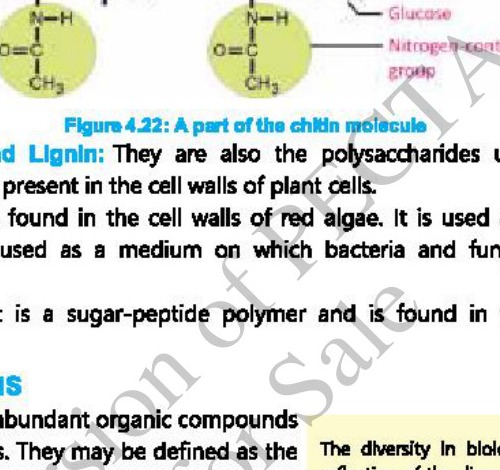
It is a modified form of cellulose. It is found in the exoskeletons of crabs, lobsters and insects. It also makes the cell wall of fungi. Like cellulose, it is also a polymer of glucose. The linkage between glucose monomers is also like that found in cellulose. However, in chitin each glucose molecule has been modified by the addition of a nitrogen-containing group. Only few organisms can digest it.
5. Pectin and Lignin: They are also the polysaccharides used as building material. They are present in the cell walls of plant cells.
6. Agar: It is found in the cell walls of red algae. It is used as a thickener in foods. It is also used as a medium on which bacteria and fungi are grown in laboratories.
7. Mucein: It is a sugar-peptide polymer and is found in the cell walls of prokaryotes.
Proteins Proteins are the polymers formed by the peptide linkage of monomers called amino acids. The most abundant organic compounds in cell. Different proteins may have a few to 3000 amino acids in their make-up (e.g., Insulin has 51 amino acids, Haemoglobin has 574 amino acids).
★ Diversity of protein: The diversity in biological world is the reflection of the diversity of structure and function that exists in proteins.
Composition of Amino acid Amino acid is the basic structural unit of proteins. It is an organic molecule, in which four groups; an amino group (NH₂), a carboxyl group (COOH), a hydrogen group (H) and a side group (R); are attached to the same carbon atom (alpha carbon).
Occurrence and types Although many different amino acids occur in nature, about 170 types of amino acids have been reported to occur in living organisms (in cells and tissues). Of these, about 25 types of amino acids may take part as building units of proteins. Most of the proteins are, however, made of 20 types of amino acids.
Identity and uniqueness The identity and unique chemical properties of each amino acid are determined by the nature of its side group (R), covalently bonded to alpha carbon. For example, R may be a hydrogen atom as in glycine, or CH₃ as in alanine, or any other group.
Essential and Non-essential Amino acids
Out of 20 amino acids, our bodies can make eleven amino acids. These are called non-essential amino acids and include alanine, arginine, asparagine, aspartic acid, cysteine, glutamic acid, glutamine, glycine, proline, serine, and tyrosine. The remaining nine amino acids cannot make our bodies on its own and must obtain these amino acids by eating various foods. These are called essential amino acids and include methionine, valine, tryptophan, isoleucine, leucine, lysine, threonine, phenylalanine and histidine (necessary only for babies).
Peptide bond and dehydration synthesis: A covalent bond that links two amino acids is known as a peptide bond. Each amino acid has an amino group at one end and a carboxyl group at the other end. When two amino acids are brought closer, dehydration synthesis occurs between the amino group of one and the carboxyl group of second amino acid. It results in the release of a molecule of water and formation of a peptide bond between "N" and "C" of adjacent amino acids.
Polypeptide chain formation A dipeptide has an amino group at one end and a carboxyl group at the other end of molecule. So, both reactive parts are available for further peptide bonds. Addition of amino acids ultimately leads to polypeptide chains. A protein is composed of one or more polypeptide chains, e.g., insulin protein contains two polypeptide chains while haemoglobin protein has four polypeptide chains. Polypeptide chains assume different shapes on the basis of number, types and sequence of amino acids. It gives different levels of structure to proteins.
The diversity of proteins ranges from simpler (consisting of linear chains of amino acids) to complex proteins (structural modifications in linear chain). The following are different levels at which proteins are built.
1. Primary Structure
• Formation: The primary structure of a protein molecule is formed by the linear arrangement of amino acids. It represents the number and sequence of amino acid molecules in a polypeptide chain.
• All protein molecules (whether simple or complex) have specific primary structures.
• The primary structure of insulin reveals that it is composed of two polypeptide chains. The smaller alpha chain has 21 amino acids while the longer beta chain is made of 30 amino acids.
★ Protein in Humans: These are over 10,000 proteins in human body and each of these has its specific primary structure i.e., specific number, specific sequence and specific types of amino acids.
• Bonds: If proteins are made up of single polypeptide chain, bonds are peptide bonds. If proteins have more than one polypeptide chains, they are attached with disulphide bond or bridge.
Importance of primary structure The number and sequence of amino acids is highly specific in the primary structure of a protein. This specificity in primary structure is determined by the order of nucleotides in DNA. Any change results in abnormal protein that fails to carry out its normal function.
Substitution of amino acids
The number, sequence and types of amino acids is highly specific in the primary structure of a protein, for its proper functioning. This specificity in primary structure is determined by the order of nucleotides in DNA. Any change results in abnormal protein that fails to carry out its normal function.
For example, sickle cell haemoglobin is formed by a mistake in the arrangement of only one amino acid in position six in each beta chain. In sickle cell haemoglobin, amino acid valine is present in the place of glutamic acid. Due to sickle cell haemoglobin, red blood cell get sickle shapes and abnormal haemoglobin cannot transport sufficient oxygen. This disease is known as sickle cell anaemia.
2. Secondary Structure
• Formation: The folding or coiling patterns that occur within a polypeptide chain is called secondary structure.
Types
• Coiling of a polypeptide chain results in alpha helix
• The folding of polypeptide makes a pleated sheet.
• Bonds: Both these structures are maintained by hydrogen bonds between amino and carboxyl groups of nearby amino acids in the chain.
3. Tertiary Structure
• Formation: When the secondary structure further folds up and gets a complicated globular shape. It is called the tertiary structure of protein. These are more complex proteins.
• Bonds: The globular shape is maintained by ionic, hydrogen and disulphide bonds. These bonds contribute to the overall stability and shape of the protein.
• Most stable tertiary structure: Amino acids in a polypeptide chain interact with water to give the most stable tertiary structure in the form of a globular shape. These are hydrophilic and hydrophobic interactions. The hydrophobic (non-polar) amino acids aggregate in such a way that they disrupt hydrogen bonding of water molecules and so are buried inside. At the same time the hydrophilic (polar) amino acids turn out, towards the surface of water.
4. Quaternary Structure
• Formation: Aggregation of two or more than two tertiary polypeptide structures is called quaternary structure.
• Bonds: When two or more polypeptide chains with tertiary structures are held together by hydrophobic interactions, hydrogen bonds and ionic bonds, they form most complex proteins.
Characteristics of Fibrous and Globular Proteins
Characteristics | Fibrous Proteins | Globular Proteins
Shape | In the form of fibrils | Spherical or ellipsoidal
Structure | Primary or Secondary | Tertiary or quaternary
Role | Structural | Functional
Crystallization | Non crystalline and elastic | Can be crystallized
Solubility | Insoluble | Soluble in salt, acid or base solutions and in aqueous alcohol
Disorganization | Do not disorganize easily | Disorganized with changes in environment
Examples | Silk fibre-from the webs of silk worm and spider
Actin in muscle cells
Fibrin – in blood clots
Keratin – in nails, hairs, beak, skin etc.
Collagen – in matrix of connective tissues | Enzymes – biocatalyst
Antibodies – active against invading antigens
Hormones – regulate body's activities
Haemoglobin – oxygen carrying protein
Proteins carry out virtually all activities of living organisms. Some of their remarkable structural and functional roles are given below.
1. Component of plasma membrane
Proteins are an important part of the composition of all plasma membranes.
2. Channel proteins: Channel proteins are the membranes of cells control the movement of materials in and out of cells. For example, proteins make sodium – potassium pump in the cell membrane of neurons. This pump controls the movement of Na⁺ and K⁺ ions in and out of nerve cell.
3. Fibrous Proteins: Some fibrous proteins e.g., collagen and keratin make almost whole structures of cartilage and hair, nails respectively.
4. Enzymes: Enzymes are a class of proteins that catalyse the metabolism of cells. They are a much diverse class of proteins. For example, proteases catalyse the breakdown of proteins, polymerases catalyse the synthesis of polymers.
5. Hormones: Some very important hormones of animals are proteins or peptides in nature. For example; insulin (controls blood glucose level), antidiuretic hormone (increases water retention by kidneys), oxytocin (regulates milk production).
6. Globular Proteins: Some globular proteins work to transport different materials throughout the body. For example; haemoglobin and myoglobin transport O₂ and some CO₂, and cytochromes work in electron transport chain as electron carriers.
7. Albumin is a blood protein that maintains osmotic concentration of blood and keeps its ability to flow.
8. Blood clotting is important to prevent the loss of blood after an injury. Fibrinogen protein is present in blood. When an injury occurs, fibrinogen is activated into fibrin. The fibrin makes fibres and a clot is formed.
9. Contractile proteins: All types of contractions in living matter are due to the actions of proteins. For example, actin and myosin are main proteins of muscles. They are responsible for muscular contractions. Tubulin protein makes spindle fibres.
10. Defence proteins: Antibodies are important proteins. They recognize and combine with foreign substances (antigens) and convert them into harmless products.
11. Ion-binding proteins: Some ion-binding proteins store ions in different parts of body. For example, ferritin is the main intracellular iron storage protein. Similarly, casein is a milk protein that stores potassium and calcium ions.
12. Repressors are the proteins that regulate gene action by preventing the synthesis of RNA. These proteins allow genes to work where and when required.
Lipids Lipids are a loosely defined group of non-polar molecules that are insoluble in water but soluble in organic solvents (e.g., ether, alcohol, etc). They are a diverse group of molecules and are classified as acylglycerols, waxes, phospholipids, terpenes, steroids and prostaglandins.
(A) Acylglycerols (Fats and Oils)
Subunits Acylglycerols are composed of two subunits: glycerol and fatty acid.
Oil The acylglycerols which are liquid at room temperature, are called oils.
Fats The acylglycerols which are solid at room temperature, are called fats. In animals, most acylglycerols are fats.
In plants, most acylglycerols are oils; for example, peanut oil, corn oil, castor oil etc.
Chemical Composition Chemically, acylglycerols are the esters of fatty acids and alcohol. They are synthesized through dehydration synthesis (H is released from alcohol and OH from an acid) as shown below.
C₂H₅OH + HOOCCH₃ ——→ C₂H₅OCOCH₃ + H₂O
alcohol acetic acid an ester
(ethylacetate)
Example The most widely found acylglycerols are triacylglycerol (triglycerides), also called neutral lipids.
Glycerol It is a 3C alcohol and each of its carbon bears a hydroxyl group. The 3 carbons of glycerol form the backbone of acylglycerol molecule, to which three fatty acids are attached.
Fatty acids These are responsible for all the characteristics of acylglycerols. Fatty acids are long hydrocarbon chains (with carbon in even number 4 – 30), ending in a carboxyl (- COOH) group. They vary in length and may be as straight chains (in animals) or branched or ringed (in plants). They are of two types:
• Saturated fatty acids contain no double bond in their hydrocarbon chain. In saturated fatty acids, all internal carbon atoms possess hydrogen side groups. These fatty acids make straight chains, and have a high melting point.
• Unsaturated fatty acids have double bonds (6 maximum) between one or more pairs of carbon atoms. The double bonds replace some of the hydrogen atoms. Therefore, unsaturated fatty acids contain fewer than the maximum number of hydrogen atoms. These fatty acids form bent chains, and have a low melting point.
★ If a fatty acid has one double bond it is called mono-unsaturated.
★ If there are more than one double bond, it is called poly-unsaturated.
Solubility Solubility of fatty acids in organic solvents and their melting points increase with increasing number of carbon atoms in their chains.
Efficient energy-storage Acylglycerols are efficient energy-storage molecules. It is due to higher number of C-H bonds in them. They are insoluble, because of their non-polar structure. Therefore, they can be deposited at specific storage locations within organism. Animal fats contain more energy than do plant oils, because they contain saturated fatty acids and so contain more C-H bonds. On the other hand, plant oils have unsaturated fatty acids and have comparatively lesser number of C-H bonds. When organisms have to store glucose for long periods, they usually convert it into fats or oils.
B. Waxes
Physical Properties Waxed are derived from acylglycerols. They have high melting points, because of large number of C atoms and so are solid at room temperature.
Chemical Composition Chemically, waxes do not have any well-defined structure and composition. They are mixtures of long chain alkanes (with carbon atoms in odd number; C25-C35), alcohols (other than glycerol), ketones and long chain fatty acids.
Significances of waxes
• Waxes are chemically inert. Like other lipids, waxes are strongly hydrophobic. So, they act as protective coverings and water barriers for living organisms.
• Waxes are widespread as protective coatings on fruits and leaves.
• Some animals like insects, birds, sheep etc. also secrete waxes over their skin.
• Waxes are used to waterproof paper and cards. Waxes are also used in wax polishes for furniture, footwear and vehicles.
• Waxes are also used to make candles.
• Waxes with coloured pigments are used in making crayons and coloured pencils.
★ Wax production: Honeybees produce waxes and use it to make six sided (hexagonal) chambers of their combs, where honey is stored. In humans, wax is secreted by glands of the outer ear canal.
C. Phospholipids
• Chemical Composition: Chemically they are the derivatives of phosphatidic acid. Phosphatidic acid is composed of one glycerol, two fatty acids and one phosphoric acid (phosphate).
• Any nitrogenous base e.g., choline, ethanolamine or serine attaches with its phosphoric acid and makes phospholipid.
• Examples: Common examples are phosphatidyl-choline (lecithin), phosphatidyl ethanolamine and phosphatidyl serine.
Components of phospholipids Phospholipids have two parts of their molecules i.e., head and tail.
(i) Head: Head is polar and contains nitrogenous base and phosphate group.
(ii) Tail: Tail is non polar and contains the two fatty acids.
Roles of Phospholipids
• Phospholipids play important structural roles in making plasma membranes. Phosphatidyl-choline forms lipid bilayer in plasma membranes.
D. Terpenes
Composition It is a very large and diverse group of lipids. All terpenes are made of isoprene units.
Structure of formula An isoprene unit is a branched unsaturated hydrocarbon chain with the formula CH₂=C(CH₃) – CH=CH₂.
Roles Terpenes form many biologically important pigments, such as chlorophyll in plants and retinal pigments in eyes.
Example Vitamin A and rubber are also terpenes.
Steroids Steroids are lipids whose carbon skeleton is bent to form four fused rings. All steroids have the same ring pattern i.e., three 6-cornered rings and one 5-cornered ring.
• Role: Cholesterol is a common steroid in animal cell membranes. Animal cells also use it for making other steroids e.g., male and female sex hormones.
• Anabolic steroids: Today, anabolic steroids are banned. Anabolic steroids can cause serious physical and mental problems e.g., deep depression, liver damage etc.
Prostaglandins
Prostaglandins are a group of lipids that are modified fatty acids, with non- polar tails attached to a five-carbon ring.
• Role: They occur in many tissues of vertebrates, where they act as local chemical messengers. Some of them stimulate smooth muscles to contract and relax; others constrict or expand the diameter of blood vessels.
• They are also involved in inflammatory response to infection. Aspirin is the inhibitor of prostaglandins.
Role of lipids in life
Sources of energy Lipids are important sources of energy (ATP). In fact, lipids are the most energy rich of all nutrients. One gram of lipids provides 9.5 kilocalories of energy. The same amount of protein provides 5.6 kilocalories while that of carbohydrate provides 4.1 kilocalories.
Components of membranes Lipids are essential components of all cellular and subcellular membranes.
Biological carriers They serve as biological carriers for the absorption of fat-soluble vitamins A, D, E and K.
Source of fatty acids Lipids are a source of fatty acids, which are essential for various metabolisms.
Mechanical cushion Lipids play a role as a mechanical cushion/support for vital body organs.
Insulation The lipids (fats) present beneath skin, insulate the body from extreme temperatures.
Steroids perform a wide range of important biological functions. For example, cholesterol is involved in the maintenance of membranes. It also helps in lipid transport. It as a precursor of vitamin D, bile acids, and steroid hormones (androgens, oestrogens), adrenal hormones and corticosteroids.
Nucleic acid Nucleic acids are the polymers of nucleotide units. There are two main types of nucleic acids i.e., deoxyribonucleic acid (DNA) and ribonucleic acid (RNA).
Occurrence DNA is found mainly in chromosomes, with small amounts in mitochondria and chloroplasts. RNA is found in nucleolus, ribosomes and cytosol.
Composition A nucleotide is made up of a nucleoside and phosphoric acid. A nucleoside is made of a nitrogen base and a pentose sugar.
a. Pentose sugars: RNA contains ribose (C₅H₁₀O₅) while DNA contains deoxyribose (C₅H₁₀O₄) as their pentoses.
b. Nitrogenous bases: There are two types of nitrogenous bases in nucleic acids i.e., pyrimidine bases and purine bases.
• Pyrimidine: Pyrimidine is a single ringed nitrogenous base. There are three pyrimidine bases in nucleic acids. Cytosine (C) is present in both DNA and RNA, thymine (T) is present only in DNA, and uracil (U) is present only in RNA.
Purine Purine is a double ring nitrogenous base. Both DNA and RNA contain two purine bases i.e., adenine (A) and guanine (G).
One nitrogenous base is attached with carbon 1 of pentose sugar and makes a nucleoside.
c. Phosphoric acid: A nucleoside develops ester linkage with a phosphoric acid and becomes nucleotide. In this ester linkage, phosphoric acid is linked with C-5 of pentose sugar. The backbone of the structure of nucleic acids is made of sugars and phosphates.
Formation of Phosphodiester Bond
In one nucleotide, phosphoric acid has an ester linkage at C-5 of pentose sugar. This phosphoric acid develops another ester linkage at C-3 of pentose sugar of another nucleotide. In this way, each phosphoric acid has two ester linkages with two pentose sugars (one at C-5 and other at C-3).
Phosphodiester The two ester linkages developed by phosphoric acid with two pentose sugars are known as phosphodiester linkage. This linkage joins two nucleotides.
Ribonucleotides The nucleotides of RNA are known as ribonucleotides.
Deoxyribonucleotides Nucleotides of DNA are known as deoxyribonucleotides. They are named after the type of nitrogenous base.
Biological importance of nucleotides
• Nucleotides also play other critical roles in the life of cell. For example; ATP is a triphosphate nucleotide of adenine. In ATP, three phosphate groups are attached with one ribose sugar. ATP is the "energy currency" of cell. It provides energy by successively detaching its two phosphate groups and changing to ADP and AMP
• Nicotinamide Adenine Dinucleotide (NAD) is a co-enzyme. It acts as a hydrogen acceptor in oxidation-reduction reactions in cell.
• James D. Watson and Francis Crick, in 1953 put forward the model of DNA.
• Watson and Crick's Model of DNA suggests the following points:
• DNA is made of two polynucleotide chains or strands.
• The two strands are coiled around each other and make a double helix.
• The double helix is like a ladder. Its poles are made of sugars and phosphate groups. Its rungs are made of nitrogenous base pairs.
• Each base pair (rung) is made of one purine (A or G) and one pyrimidine (C or T) base.
• Two strands are held together by weak hydrogen bonds between their bases.
• Adenine in one chain makes hydrogen bonds with thymine in second chain, or vice versa. Guanine in one chain makes hydrogen bonds with cytosine in second chain, or vice versa.
• There are two hydrogen bonds between A and T pair and three hydrogen bonds between G and C pair.
• Two strands are not in the same direction with respect to their phosphodiester linkages, but are anti-parallel to each other.
Location DNA is the fundamental part of chromosomes and so is located inside nucleus in eukaryotes. As there is distinct nucleus in prokaryotes, their DNA is present in cytoplasm. In viruses, DNA is located as a core molecule, covered by a protein coat.
Function DNA is the hereditary material for all organisms (except some viruses). DNA contains the "program" that ultimately directs all cellular activities. The program in DNA is in the form of genes.
Gene A gene is a sequence of nucleotides of DNA, which codes for the formation of a polypeptide.
When a gene is turned "ON", the sequence of DNA nucleotides is transcribed into RNA and then translated into specific proteins.
In this way DNA controls the properties and activities of a cell.
★ Genetic composition of E.coli: In the chromosome of bacterium E.coli, each strand of DNA contains about 5 million bases arranged in a particular linear order. It has genes, each consisting of several hundred bases.
Ribonucleic Acid RNA
• It is composed by ribonucleotides.
• RNA is synthesized by joining ribonucleotides in front of deoxyribonucleotides of DNA by transcription process.
Types All living cells contain three types of RNA.
1. Messenger RNA (mRNA)
It consists of a single strand of ribonucleotides. Its sequence of nucleotides is complimentary to the sequence of nucleotides of one of the strands of DNA. mRNA is about 3-4% of the total amount of RNA in cell. It carries the genetic message of DNA to ribosomes to form particular protein.
2. Transfer RNA (tRNA)
It is comparatively small. It is a helical structure and its molecule resembles a clover leaf. It consists of 10-15% of the total amount of RNA in cell. tRNAs transport amino acids to ribosome and mRNA, in the process of protein synthesis.
3. Ribosomal RNA (rRNA)
It is synthesized by the DNA of nucleoli. After its synthesis, ribosomal RNA is joined with ribosomal protein and ribosomes are formed. It comprises about 80% of the total RNA in cell. rRNA acts as the machinery for synthesis of proteins in ribosomes.
Central Dogma
All organisms use the same basic mechanism of reading and expressing genes, which is often referred to as central dogma.
Transcription The first step of central dogma is the transfer of information from DNA to RNA, which occurs when an RNA copy of the gene is produced. The process is called transcription. In prokaryotes it occurs in the cytoplasm while in eukaryotes transcription occurs in the nucleus. Transcription requires a promotor sequence to be started.
Translation The second step of the central dogma is the transfer of information from RNA to proteins, which occurs when the information contained in the RNA is used to direct the synthesis of proteins. This process is called translation. In prokaryotes translation directly starts in the cytoplasm as they lack nucleus, while in eukaryotes mRNA has to be transported to the cytoplasm for decoding and protein synthesis. Translation starts from a start codon which codes for Methionine.
In this way DNA controls the properties and activities of a cell.
Conjugated molecules are formed by the combination of two or more molecules belonging to different categories.
Examples Some important conjugated molecules are as follows.
(i) Glycoproteins: They are formed by covalent linkage between a protein and a carbohydrate polymer. They occur widely in nature as integral structural component of membranes; in blood serum; as cellular secretions; and in cartilage, eyes, skin etc.
(ii) Glycolipids: They are formed by a covalent linkage between a lipid and a carbohydrate. They are an integral structural component of membranes.
(iii) Lipoproteins: They are a class of biomolecules which are formed by hydrophobic interactions (not covalent or ionic bonds) between lipids and proteins. Lipoproteins are the basic structural framework of all types of plasma membranes. Lipids are transported in blood as very low-density lipoproteins.
(iv) Nucleoproteins: They are formed by ionic bonds between chromosomal DNA and proteins. For example, histone proteins are bound to DNA to form nucleosomes. They stabilize chromosomal structure in eukaryotes and also play an important role in the regulation of gene expression.
More figures from this unit